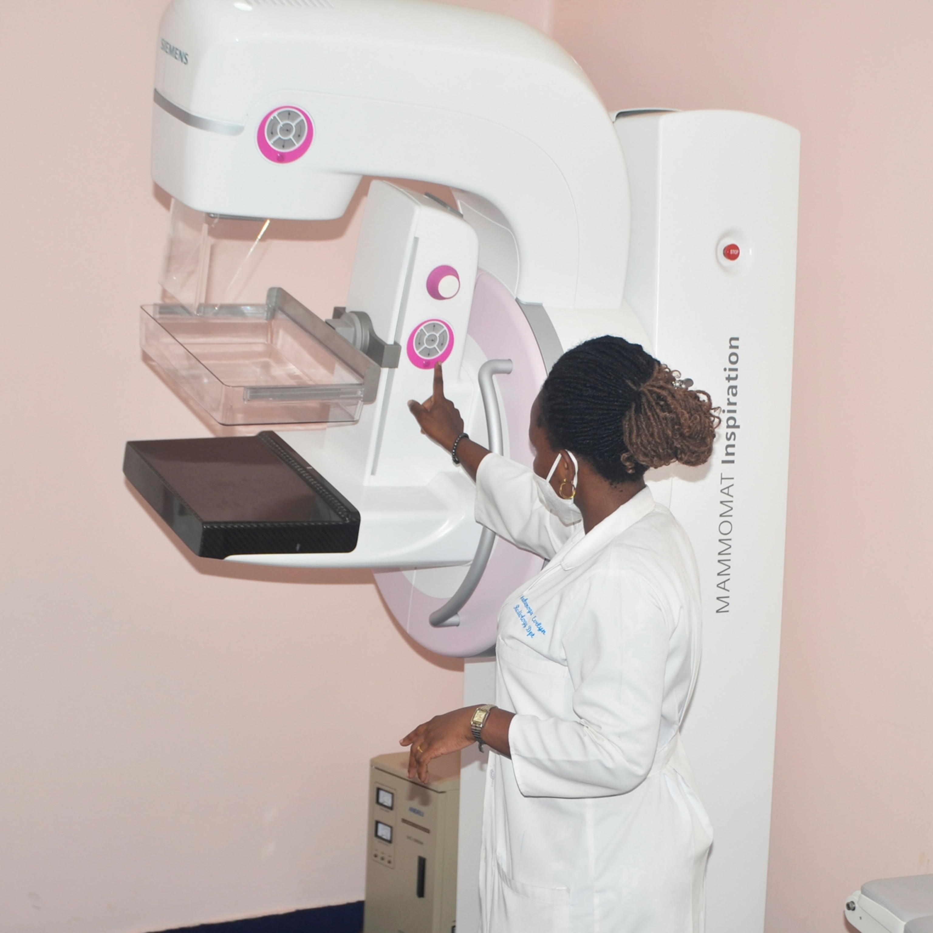
What are dense breasts? How this common condition complicates cancer detection
If you’ve been told you have dense breasts, your mammogram results might not tell the full story. Here’s how breast density affects cancer detection—and which extra screenings can help.

After receiving her routine mammogram results, 46-year-old Nicole Pearl found herself in what she calls “no man’s land.” The letter from her doctor said she was “clear” of cancer, but also mentioned she had dense breasts—leaving her uncertain about what that meant for her health. “So, I may not actually be clear and have the option for more testing,” she says.
Pearl’s experience is far from unique. According to Memorial Sloan Kettering Cancer Center, around 40 percent of women have mostly dense breasts, and 10 percent have extremely dense breasts. Younger women are more likely to have dense breast tissue, although it typically decreases with age.
But the issue isn’t dense breasts themselves; it’s how they can obscure cancer on mammograms. Both cancer and dense tissue show up white, making it harder to detect abnormalities. While dense breasts are linked to a higher cancer risk, the exact risk depends on other factors. Here’s what you need to know.
What are dense breasts?
Dense breasts aren’t larger, lumpier, or smoother than average, says Richard Reitherman, medical director of breast imaging at Orange Coast Medical Center in Fountain Valley, California.
Instead, they have more fibrous and glandular tissue compared to fatty tissue, which makes it harder to detect cancer on mammograms.
Reitherman says there are four levels of breast density—A, B, C, and D. The top two levels (C and D) are considered “dense” and are the most concerning. “You can’t see much at all on a mammogram,” he says.
(Do increased breast cancer screenings save lives? Doctors can’t agree.)
Research across 29 studies shows combining mammograms with ultrasound increases cancer detection in women with dense breasts by 40 percent. However, mammograms still matter greatly for all women because they can detect “alterations and distortion” that ultrasounds can’t see, Reitherman says.
How do I know if I have dense breasts?
You can’t determine breast density just by feel or appearance—it requires diagnostic testing. And “you can’t tell by age,” says Laurie Margolies, vice chair of breast imaging at Mount Sinai Health System in New York.
“Breast density is very often related to your [genetics],” she says. Women with a lower body mass index (BMI) are more likely to have dense breasts, but this isn’t always the case, she adds. ”The only way you know for sure is mammography, or occasionally a chest CT,” says Margolies.
(New research shows how genes can identify risks of breast cancer.)
In March 2023, the U.S. Food & Drug Administration (FDA) updated the Mammography Quality Standards Act (MQSA) to require that all mammography reports include information about breast density, ensuring that patients are informed about this important aspect of their results. This regulation is set to take full effect by September 2024. However, the challenge for many women comes after receiving this information—what should they do next?
What should you do if you have dense breasts?
While dense breasts can make it harder to spot cancer, they don’t mean you have cancer. In fact, a 2023 study found that women with dense breasts often have an inaccurate “perceived personal” risk of breast cancer, leading to overestimating their risk of breast cancer.
But, “you can do more than a mammogram for early detection,” says Margolies, who suggests discussing options like ultrasound or MRI with your doctor. She adds that for every thousand women with dense breasts, additional screenings with ultrasound can uncover about three more cancers, while MRI could detect up to 16 more.
(When should you get screened for breast cancer—and how often?)
So why aren’t patients with dense breasts just routinely scheduled for additional testing? “The reason we can’t do that is because we’re only seeing one part of you, your mammogram,” says Kemi Babagbemi, a board-certified radiologist specializing in Women’s Imaging at Weill Cornell Medicine in New York.
Some places may not routinely offer or recommend MRIs unless other risk factors are present, as these tests are more expensive and can lead to false positives. “Your primary provider needs to be doing a risk assessment to determine what other risk factors for breast cancer you have to go to the next step,” Babagbemi adds.
Who are most at risk?
It’s scary to hear stats of increasing breast cancer rates and to wonder if your testing was correct. But, Reitherman says that there are steps you can even take on your own to determine your risk, in addition to, not in place of, a mammogram.
For younger women under 40, breast cancers tend to be more aggressive, so it’s important for younger women to fully understand their breast densities and advocate for proper testing. Black women, who are statistically more likely to die from breast cancer, along with other high-risk groups, should also be particularly mindful of their results.
“I encourage any woman wanting to explore her risk of breast cancer, download each of these models, put in the requested information, and compare the outcomes,” says Reitherman. He suggests tools like the IBIS Breast Cancer Evaluation Tool, the ACSC Advanced Breast Cancer Risk Calculator, and the National Cancer Institute’s Online Calculator. It’s also helpful to review these results with your healthcare provider, he adds.
Finally, experts agree on one key point: advocate for yourself. Ask questions, push for additional testing if needed, and don’t skip your annual screenings.


